- News ›
- Understanding WHO Brain Tumour Classifications
Understanding WHO Brain Tumour Classifications
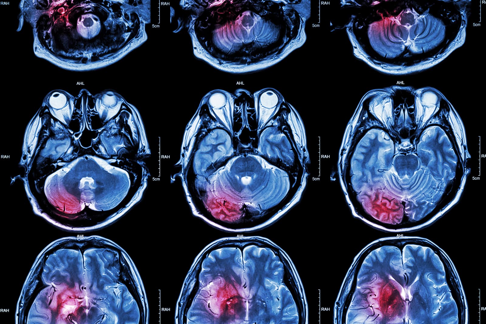
When someone is first told they have a brain tumour, one of the most important concepts they encounter is the WHO classification system. This framework, developed by the World Health Organization, is used worldwide to describe and grade brain tumours.
It helps specialist teams understand how a tumour behaves, how it may develop over time, and which treatment pathways may be most appropriate. Here is a guide to WHO classification and the role it plays in decision-making about neurological conditions.
It is not based solely on where a tumour is located, but on how the tumour cells look under the microscope, their genetic features, and their likely behaviour. This approach allows clinicians to develop a more detailed understanding of each individual diagnosis.
Modern classifications increasingly integrate molecular and genomic information alongside traditional histology. This shift has significantly improved diagnostic accuracy and supports more personalised treatment planning.
It is important to understand that grading is not a prediction of outcome for an individual, but a framework used to guide clinical decision-making.
For example, tumours that may appear similar under a microscope can behave very differently depending on their genetic profile. This is particularly relevant in conditions such as:
This integration of molecular data allows clinicians to move towards truly personalised treatment strategies rather than relying on imaging or histology alone.
Many are slow-growing, but classification helps determine whether active monitoring or targeted treatment may be appropriate.
This integrated approach ensures that every case is considered individually, with a focus on precision and clarity.
Learn more about advanced diagnostics and personalised treatment pathways for complex brain tumours at Queen Square Neuroscience Health Centre.
It helps specialist teams understand how a tumour behaves, how it may develop over time, and which treatment pathways may be most appropriate. Here is a guide to WHO classification and the role it plays in decision-making about neurological conditions.
What is the WHO classification system?
The WHO classification system is a globally recognised method for categorising brain and spinal tumours based on their biological characteristics.It is not based solely on where a tumour is located, but on how the tumour cells look under the microscope, their genetic features, and their likely behaviour. This approach allows clinicians to develop a more detailed understanding of each individual diagnosis.
Modern classifications increasingly integrate molecular and genomic information alongside traditional histology. This shift has significantly improved diagnostic accuracy and supports more personalised treatment planning.
WHO brain tumour grades: an overview
Brain tumours are commonly assigned a WHO grade from I to IV. These grades describe how the tumour cells appear and behave, particularly in terms of growth patterns and cellular activity.WHO Grade I
These tumours tend to grow slowly and have well-defined boundaries. They are often more straightforward to manage and may be suitable for long-term monitoring or targeted treatment depending on location and symptoms.WHO Grade II
These tumours are typically slow-growing but can infiltrate surrounding brain tissue. Over time, they may show changes in behaviour, which is why careful long-term follow-up is essential.WHO Grade III
These tumours show more active cellular growth and are considered more aggressive in behaviour than lower-grade tumours. They require structured, specialist-led management and ongoing assessment.WHO Grade IV
These are the most biologically active tumours, characterised by rapid growth and complex cellular patterns. They require highly coordinated care involving advanced imaging, genomic testing, and specialist treatment planning.It is important to understand that grading is not a prediction of outcome for an individual, but a framework used to guide clinical decision-making.
Why molecular classification matters for brain tumour diagnosis
In modern neuro-oncology, WHO classification goes beyond grading alone. Molecular and genetic markers now play a key role in defining tumour types more precisely.For example, tumours that may appear similar under a microscope can behave very differently depending on their genetic profile. This is particularly relevant in conditions such as:
- Astrocytoma – where genetic changes help determine tumour subtype and behaviour
- Oligodendroglioma – often defined by specific genetic markers such as 1p/19q co-deletion
- Glioblastoma – where molecular features help guide treatment pathways and clinical planning
This integration of molecular data allows clinicians to move towards truly personalised treatment strategies rather than relying on imaging or histology alone.
Common tumour types and WHO classification
WHO classification is applied across a wide range of brain and cranial conditions, including both primary brain tumours and related neurological growths.Gliomas
Gliomas arise from supporting brain cells and include astrocytoma, oligodendroglioma, and glioblastoma. These are classified based on both grade and molecular profile, which helps guide advanced treatment planning and monitoring strategies.Meningioma
Meningiomas arise from the membranes surrounding the brain and spinal cord. They are graded from I to III depending on cellular behaviour.Many are slow-growing, but classification helps determine whether active monitoring or targeted treatment may be appropriate.
Vestibular schwannoma (acoustic neuroma)
Although not graded in the same way as gliomas, vestibular schwannomas are still evaluated within a structured diagnostic framework to guide management decisions, particularly when considering hearing preservation and balance function.Brain metastases
Secondary brain tumours are assessed differently, as classification focuses on the primary cancer type and the pattern of spread to the brain. Imaging and molecular data help determine the most appropriate targeted approach.How WHO classification supports brain tumour treatment planning
One of the key strengths of the WHO system is its role in treatment planning for brain tumours. At a specialist centre such as Queen Square Neuroscience Health Centre, classification is not viewed in isolation. Instead, it is combined with:- High-resolution MRI and advanced imaging techniques
- Genomic and molecular testing
- Neurological assessment and symptom evaluation
- Multidisciplinary team review
- Specialist neurosurgical and radiosurgical input, including Gamma Knife where appropriate
This integrated approach ensures that every case is considered individually, with a focus on precision and clarity.
Why brain tumour classification is continually evolving
WHO classification is not static. It is regularly updated as scientific understanding improves. Advances in molecular biology, imaging technology, and clinical research continue to refine how tumours are defined and grouped.Learn more about advanced diagnostics and personalised treatment pathways for complex brain tumours at Queen Square Neuroscience Health Centre.